

Autism and Sleep – Research Updates
Dara S. Manoach, Ph.D., discusses research updates on autism and sleep. She outlines aspects of a good night’s sleep and emphasizes the role of sleep cycles in cognition, memory, and learning. The speaker considers how to treat neurodevelopmental disorders by targeting sleep spindle rhythms which affect thalamocortical neuronal loops, essential to memory and attention. Manoach outlines how spindle rhythms correlate to memory consolidation during sleep in schizophrenia and autism, and highlights technological advancements that allow for broader sample sizes and more accessible studies. The presenter notes potential treatment goals and ongoing research before the Q&A.
A good night’s sleep
Manoach explains that a night’s sleep is divided into REM and non-REM cycles, with non-REM further divided into three stages based on slow-wave activity. The speaker’s research focuses on non-REM Stage 2 sleep, which occurs in the early morning and is characterized by sleep spindle brain rhythms (1:15). Manoach considers how sleep affects cognition, highlighting that critical aspects of learning and memory occur during sleep (2:15). She cites ancient references to the interaction of memory and sleep (Quintilianus, 95 AD) and highlights a 2001 study that kicked off the current explosion in sleep and memory research (3:10).
- Sleep, Learning, and Dreams: Off-line Memory Reprocessing (Stickgold et al., 2001)
Sleep, memory, and learning
The presenter cites studies showing that sleep prioritizes memory for emotional information and helps extract the main point of an idea or experience. Sleep also increases spatial navigation and facilitates language learning in infants (4:25). The speaker asserts that this body of work has led to a paradigm shift in understanding sleep as an active period of cognitive functioning that facilitates learning and memory (6:00).
- The role of sleep in declarative memory consolidation: passive, permissive, active or none? (Ellenbogen et al., 2006)
- Sleep Preferentially Enhances Memory for Emotional Components of Scenes (Payne et al., 2008)
- Dreaming of a Learning Task Is Associated with Enhanced Sleep-Dependent Memory Consolidation (Wamsley et al., 2010)
- Sleep inspires insight (Wagner et al., 2004)
- The Sleeping Infant Brain Anticipates Development (Friedrich et al., 2017)
The speaker describes finger-tapping motor sequence tasks (MST) in which participants use a keyboard to type a number sequence as quickly and accurately as possible. The following day, they complete the MST again to test learning improvements after a night of sleep (6:40). Research shows significant improvement in MST results after 12-hour intervals with a sleep sequence (7:45). Time of MST training (10 AM vs 10 PM) did not affect following day outcomes, and a 12-hour interval with no sleep cycle did not yield performance improvements (8:15). These findings, Manaoch asserts, emphasize the critical role of sleep in memory and learning.
- The acquisition of skilled motor performance: Fast and slow experience-driven changes in primary motor cortex (Karni et al., 1998)
- Practice with Sleep Makes Perfect (Walker et al., 2002)
Sleep spindles and memory consolidation
MST studies in people with schizophrenia show a normal amount of learning during training. However, after a night of sleep, MST performance did not significantly improve, meaning that individuals with schizophrenia do not get the same benefits from sleep as their peers (9:23). The speaker notes that this failure of overnight improvement occurred in the context of significantly reduced sleep spindles, which are a critical mechanism of memory consolidation (10:00). Manoach states that memory consolidation deficits are also characteristics of autism, though the link has been better established in schizophrenia literature (12:17).
The presenter outlines her research model, highlighting the importance of forging empirical links in causal chains across genes, circuits, physiology, cognition, diagnosis, and treatment (13:04). She describes brain spindles as a rhythm of Stage 2 non-REM sleep, which is recorded on an EEG as one-to-two-second bursts of brain activity. Spindles are initiated in the thalamus and sent to the cortex via thalamocortical neuron loops (e.g., bursts of brain activity), regulated by the thalamic reticular nucleus (TRN). The TRN is essential for sensory gating, or filtering out sensory inputs. Manoach asserts that spindle deficits in schizophrenia suggest a problem in this circuitry related explicitly to TRN modulation (12:50).
- A failure of sleep-dependent procedural learning in chronic, medicated schizophrenia (Manoach et al., 2004)
- Reduced Sleep Spindles and Spindle Coherence in Schizophrenia: Mechanisms of Impaired Memory Consolidation? (Wamsley et al., 2012)
She cites research showing that dysfunctional thalamocortical circuitry can lead to attention fragmentation and impaired sensory gating in schizophrenia (14:30). Studies also show hyperconnectivity between the thalamus and motor sensory cortex associated with spindle deficits. Therefore, she asserts that research to date suggests decreased spindles and increased connectivity reflect the same thalamocortical circuit dysfunction in schizophrenia (16:00).
- The thalamic reticular nucleus: structure, function, and concept (Pinault, 2004)
- Oxidative stress-driven parvalbumin interneuron impairment as a common mechanism in models of schizophrenia (Stuellet et al., 2017).
- Disorders of attention and perception in early schizophrenia (McGhie & Chapman, 1961)
- Translational utility of rodent hippocampal auditory gating in schizophrenia research: a review and evaluation (Smucny et al., 2015)
- Increased Thalamocortical Connectivity in Schizophrenia Correlates With Sleep Spindle Deficits: Evidence for a Common Pathophysiology (Baran et al., 2019)
Genetic underpinnings
Manoach cites two animal model studies that found spindle deficits in mice with schizophrenia risk gene CACNA1I. Spindle deficits were correlated with reductions in thalamocortical circuitry (17:30). The speaker explains that autism risk genes, like PTCHD1, are also implicated in TRN dysfunction and have been correlated with spindle reduction and reduced visual gating leading to fractured attention in animal models. These data, she asserts, directly link gating issues to attention deficits and spindle dysfunction (19:40). She explains how TRN mutations in early development can predispose individuals to neurodevelopmental conditions like autism (21:15).
- A rare schizophrenia risk variant of CACNA1I disrupts CaV3.3 channel activity (Andrade et al., 2016)
- Effects of a patient-derived de novo coding alteration of CACNA1I in mice connect a schizophrenia risk gene with sleep spindle deficits (Goshal et al., 2020)
- Thalamic reticular impairment underlies attention deficit in Ptchd1Y/− mice (Wells et al., 2016)
- New views of the thalamic reticular nucleus in the adult and the developing brain (Mitrofanis & Guillery, 1993)
- Early motor activity drives spindle bursts in the developing somatosensory cortex (Khazipov et al., 2004)
Sleep in autism
The presenter explains that 80% of autistic children experience sleep disturbances, which can exacerbate symptoms and impact daily function. She outlines a recent home-sleep EEG study that revealed autistic people had worse sleep quality than their non-autistic counterparts, specifically related to sleep latency, sleep efficiency, night wakings/arousals, and percentage of time in non-REM Stage 1 sleep (22:20). The same study also found reduced spindle rates in Stage 3 non-REM sleep and reduced thalamocortical coordination, with later and more variable spindle peaks compared to controls (24:00). These data, Manoach explains, are consistent with disruption to TRN during sleep. She hypothesizes that, as in schizophrenia, there is less gating of auditory information due to TRN deficits in autism (26:00).
- Dyscoordination of non-rapid eye movement sleep oscillations in autism spectrum disorder (Mylonas et al., 2022)
- Increased resting-state thalamocortical functional connectivity in children and young adults with autism spectrum disorder (Baran et al., 2022)
- Sensory experiences of children with autism spectrum disorder: In their own words (Kirby et al., 2014)
Sleep-dependent memory consolidation in autism
The presenter notes that MST studies in autism do not report deficits in sleep-dependent memory consolidation. Where typically developing individuals had normal spindle count, autistic participants showed an inverse number comparatively (27:00). Manoach considers the role of spindles if not memory. She posits that they may support overall sleep cycles and, therefore, spindle deficits may contribute to sleep disruption in autism (28:00).
- Dyscoordination of non-rapid eye movement sleep oscillations in autism spectrum disorder (Mylonas et al., 2022)
Ongoing work
Manaoch’s ongoing work looks at the links between sleep disruption in autism and spindle deficits, circadian disruptions, thalamocortical circuit function (gating), EEG abnormalities, and relations to sensory sensitivities. She notes a pilot study on the physiological effects of melatonin (29:00). The speaker considers the issue of small sample sizes in sleep analysis and explains how they are scaling up research with a wearable sleep headband that can be used at home, making sleep studies more accessible (30:00).
She outlines part of her lab’s work with closed-loop auditory stimulation (CLAS) during a nap, monitored by EEG (31:45). Results show that CLAS has a strong effect on slow-wave and sigma rhythms, but it did not lead to better memory in this study. However, researchers now posit that timing matters (32:45). Manoach explains that the closer CLAS occurs to a sleep upstate, the more likely it is to evoke a slow-wave and spindle creation (33:30).
- The effects of closed-loop auditory stimulation on sleep oscillatory dynamics in relation to motor procedural memory consolidation Get access Arrow (Baxter et al., 2023)
Accessibility and large sample sizes
The speaker reiterates the usability of the new sleep headbands and discusses their ongoing piloting process spanning the entire U.S. (35:15). She notes experiment preparation, procedures, and support, emphasizing the accessibility of participating from home (35:55). Preliminary data show no spindle deficits in non-REM Stage 2 or Stage 3 as a whole. However, when assessed by sex, spindle deficits are constrained to males. Manaoch considers whether other inconclusive studies have considered sex and how results may differ if they had (36:30). She notes that spindle deficits have been observed in studies with only male participants and speaks to the importance of large, diverse sample sizes. The presenter notes that study protocols and tools have been adapted for those with profound autism, citing the lack of studies in this group and the need for broader samples (37:50). She briefly describes ongoing sleep biomarker studies and outlines potential treatment goals (40:25):
- Correct thalamocortical circuit function
- Increase spindles or improve coordination
- Improve sensory gating
- Restore sleep
- Support the daily life of those with autism and their caregivers
Manoach thanks and acknowledges her research team and funders (41:40). She highlights that her lab is actively recruiting participants for one of the sleep studies discussed and provides her contact information before the Q&A (43:15).
To participate in the study mentioned in the presentation, see: Participate in our Research – Sleep, Cognition and Neuropsychiatry (SCAN) Lab

Dara S. Manoach, PhD, Paul B. and Sandra M. Edgerley MGH Research Scholar 2019-2024 is a Professor of Psychology in the Department of Psychiatry at Harvard Medical School. She is a neuropsychologist who received her Ph.D. in psychology from Harvard University. She completed a clinical psychology internship at McLean Hospital and a fellowship in clinical neuropsychology at the Behavioral Neurology Unit of Beth Israel Hospital. She is in the Department of Psychiatry at Massachusetts General Hospital and is a Faculty Associate at the Lurie Center for Autism. She is based at the Athinoula A. Martinos Center for Biomedical Imaging where she directs the Sleep, Cognition, and Neuropsychiatry (SCAN) Lab.
1:15 – A good night’s sleep
4:15 – The role of sleep in memory and learning
6:40 – Motor sequence tasks (MST)
9:23 – Sleep spindles and memory consolidation
14:30 – Thalamocortical circuitry
17:30 – Genetic underpinnings
22:20 – Sleep in autism
27:00 – Sleep-dependent memory consolidation in autism
29:00 – Ongoing work
35:15 – Accessibility and sample size
40:25 – Treatment goals
43:15 – Q&A
Resources
Presentation Handouts
Slides in PDF
Sleep, Cognition and Neuropsychology (SCAN) Lab
Harvard studies open for enrollment
Sleep Issues Free Online App
App to identify care strategies for common sleep issue in autism
Cited Studies
Those inclined to relish in scientific controversy will not be disappointed by the literature on the effects of sleep on memory. Opinions abound. Yet refinements in the experimental study of these complex processes of sleep and memory are bringing this fascinating relationship into sharper focus. A longstanding position contends that sleep passively protects memories by temporarily sheltering them from interference, thus providing precious little benefit for memory. But recent evidence is unmasking a more substantial and long-lasting benefit of sleep for declarative memories. Although the precise causal mechanisms within sleep that result in memory consolidation remain elusive, recent evidence leads us to conclude that unique neurobiological processes within sleep actively enhance declarative memories.
Central aspects of emotional experiences are often well remembered at the expense of background details. Previous studies of such memory trade-offs have focused on memory after brief delays, but little is known about how these components of emotional memories change over time. We investigated the evolution of memory for negative scenes across 30 min, 12 daytime hours spent awake, and 12 nighttime hours including sleep. After 30 min, negative objects were well remembered at the expense of information about their backgrounds. Time spent awake led to forgetting of the entire negative scene, with memories of objects and their backgrounds decaying at similar rates. Sleep, in contrast, led to a preservation of memories of negative objects, but not their backgrounds, a result suggesting that the two components undergo differential processing during sleep. Memory for a negative scene develops differentially across time delays containing sleep and wake, with sleep selectively consolidating those aspects of memory that are of greatest value to the organism.
It is now well established that postlearning sleep is beneficial for human memory performance [1–5]. Meanwhile, human and animal studies have demonstrated that learning-related neural activity is re-expressed during posttraining nonrapid eye movement (NREM) sleep [6–9]. NREM sleep processes appear to be particularly beneficial for hippocampus-dependent forms of memory [1–3, 10]. These observations suggest that learning triggers the reactivation and reorganization of memory traces during sleep, a systems-level process that in turn enhances behavioral performance. Here, we hypothesized that dreaming about a learning experience during NREM sleep would be associated with improved performance on a hippocampus-dependent spatial memory task. Subjects were trained on a virtual navigation task and then retested on the same task 5 hr after initial training. Improved performance at retest was strongly associated with task-related dream imagery during an intervening afternoon nap. Task-related thoughts during wakefulness, in contrast, did not predict improved performance. These observations suggest that sleep-dependent memory consolidation in humans is facilitated by the offline reactivation of recently formed memories, and furthermore that dream experiences reflect this memory processing. That similar effects were not observed during wakefulness suggests that these mnemonic processes are specific to the sleep state.
Insight denotes a mental restructuring that leads to a sudden gain of explicit knowledge allowing qualitatively changed behaviour1,2. Anecdotal reports on scientific discovery suggest that pivotal insights can be gained through sleep3. Sleep consolidates recent memories4,5,6 and, concomitantly, could allow insight by changing their representational structure. Here we show a facilitating role of sleep in a process of insight. Subjects performed a cognitive task requiring the learning of stimulus–response sequences, in which they improved gradually by increasing response speed across task blocks. However, they could also improve abruptly after gaining insight into a hidden abstract rule underlying all sequences. Initial training establishing a task representation was followed by 8 h of nocturnal sleep, nocturnal wakefulness, or daytime wakefulness. At subsequent retesting, more than twice as many subjects gained insight into the hidden rule after sleep as after wakefulness, regardless of time of day. Sleep did not enhance insight in the absence of initial training. A characteristic antecedent of sleep-related insight was revealed in a slowing of reaction times across sleep. We conclude that sleep, by restructuring new memory representations, facilitates extraction of explicit knowledge and insightful behaviour.
From the age of 3 months, infants learn relations between objects and co-occurring words [1]. These very first representations of object-word pairings in infant memory are considered as non-symbolic proto-words comprising specific visual-auditory associations that can already be formed in the first months of life [2–5]. Genuine words that refer to semantic long-term memory have not been evidenced prior to 9 months of age [6–9]. Sleep is known to facilitate the reorganization of memories [9–14], but its impact on the perceptual-to-semantic trend in early development is unknown. Here we explored the formation of word meanings in 6- to 8-month-old infants and its reorganization during the course of sleep. Infants were exposed to new words as labels for new object categories. In the memory test about an hour later, generalization to novel category exemplars was tested. In infants who took a short nap during the retention period, a brain response of 3-month-olds [1] was observed, indicating generalizations based on early developing perceptual-associative memory. In those infants who napped longer, a semantic priming effect [15, 16] usually found later in development [17–19] revealed the formation of genuine words. The perceptual-to-semantic shift in memory was related to the duration of sleep stage 2 and to locally increased sleep spindle activity. The finding that, after the massed presentation of several labeled category exemplars, sleep enabled even 6-month-olds to create semantic long-term memory clearly challenges the notion that immature brain structures are responsible for the typically slower lexical development.
Behavioral and neurophysiological studies suggest that skill learning can be mediated by discrete, experience-driven changes within specific neural representations subserving the performance of the trained task. We have shown that a few minutes of daily practice on a sequential finger opposition task induced large, incremental performance gains over a few weeks of training. These gains did not generalize to the contralateral hand nor to a matched sequence of identical component movements, suggesting that a lateralized representation of the learned sequence of movements evolved through practice. This interpretation was supported by functional MRI data showing that a more extensive representation of the trained sequence emerged in primary motor cortex after 3 weeks of training. The imaging data, however, also indicated important changes occurring in primary motor cortex during the initial scanning sessions, which we proposed may reflect the setting up of a task-specific motor processing routine. Here we provide behavioral and functional MRI data on experience-dependent changes induced by a limited amount of repetitions within the first imaging session. We show that this limited training experience can be sufficient to trigger performance gains that require time to become evident. We propose that skilled motor performance is acquired in several stages: “fast” learning, an initial, within-session improvement phase, followed by a period of consolidation of several hours duration, and then “slow” learning, consisting of delayed, incremental gains in performance emerging after continued practice. This time course may reflect basic mechanisms of neuronal plasticity in the adult brain that subserve the acquisition and retention of many different skills.
Improvement in motor skill performance is known to continue for at least 24 hr following training, yet the relative contributions of time spent awake and asleep are unknown. Here we provide evidence that a night of sleep results in a 20% increase in motor speed without loss of accuracy, while an equivalent period of time during wake provides no significant benefit. Furthermore, a significant correlation exists between the improved performance overnight and the amount of stage 2 NREM sleep, particularly late in the night. This finding of sleep-dependent motor skill improvement may have important implications for the efficient learning of all skilled actions in humans.
Schizophrenia patients have difficulty mastering even rote procedural tasks in rehabilitation settings. Although most studies demonstrate intact procedural learning in schizophrenia, recent findings demonstrate that a critical component of procedural learning is dependent on sleep. This study tested the hypothesis that patients with schizophrenia have a deficit in sleep-dependent procedural learning. Using a simple, well-characterized test of motor skill learning, the finger tapping motor sequence task (MST), 26 patients with chronic, medicated schizophrenia and 14 demographically matched healthy control subjects were tested on two occasions, 24 hours apart. The main outcome measures were learning of the MST on day 1 (practice-dependent learning) and overnight, sleep-dependent improvement in performance.
Sleep spindles are thought to induce synaptic changes and thereby contribute to memory consolidation during sleep. Patients with schizophrenia show dramatic reductions of both spindles and sleep-dependent memory consolidation, which may be causally related. To examine the relations of sleep spindle activity to sleep-dependent consolidation of motor procedural memory, 21 chronic, medicated schizophrenia outpatients and 17 healthy volunteers underwent polysomnography on two consecutive nights. On the second night, participants were trained on the finger-tapping motor sequence task (MST) at bedtime and tested the following morning. The number, density, frequency, duration, amplitude, spectral content, and coherence of stage 2 sleep spindles were compared between groups and examined in relation to overnight changes in MST performance.
On the basis of theoretical, anatomical, psychological and physiological considerations, Francis Crick (1984) proposed that, during selective attention, the thalamic reticular nucleus (TRN) controls the internal attentional searchlight that simultaneously highlights all the neural circuits called on by the object of attention. In other words, he submitted that during either perception, or the preparation and execution of any cognitive and/or motor task, the TRN sets all the corresponding thalamocortical (TC) circuits in motion. Over the last two decades, behavioural, electrophysiological, anatomical and neurochemical findings have been accumulating, supporting the complex nature of the TRN and raising questions about the validity of this speculative hypothesis. Indeed, our knowledge of the actual functioning of the TRN is still sprinkled with unresolved questions. Therefore, the time has come to join forces and discuss some recent cellular and network findings concerning this diencephalic GABAergic structure, which plays important roles during various states of consciousness. On the whole, the present critical survey emphasizes the TRN’s complexity, and provides arguments combining anatomy, physiology and cognitive psychology.
Parvalbumin inhibitory interneurons (PVIs) are crucial for maintaining proper excitatory/inhibitory balance and high-frequency neuronal synchronization. Their activity supports critical developmental trajectories, sensory and cognitive processing, and social behavior. Despite heterogeneity in the etiology across schizophrenia and autism spectrum disorder, PVI circuits are altered in these psychiatric disorders. Identifying mechanism(s) underlying PVI deficits is essential to establish treatments targeting in particular cognition. On the basis of published and new data, we propose oxidative stress as a common pathological mechanism leading to PVI impairment in schizophrenia and some forms of autism. A series of animal models carrying genetic and/or environmental risks relevant to diverse etiological aspects of these disorders show PVI deficits to be all accompanied by oxidative stress in the anterior cingulate cortex. Specifically, oxidative stress is negatively correlated with the integrity of PVIs and the extracellular perineuronal net enwrapping these interneurons. Oxidative stress may result from dysregulation of systems typically affected in schizophrenia, including glutamatergic, dopaminergic, immune and antioxidant signaling. As convergent end point, redox dysregulation has successfully been targeted to protect PVIs with antioxidants/redox regulators across several animal models. This opens up new perspectives for the use of antioxidant treatments to be applied to at-risk individuals, in close temporal proximity to environmental impacts known to induce oxidative stress.
Impaired gating of the auditory evoked P50 potential is one of the most pharmacologically well-characterized features of schizophrenia. This deficit is most commonly modeled in rodents by implanted electrode recordings from the hippocampus of the rodent analog of the P50, the P20–N40. The validity and effectiveness of this tool, however, has not been systematically reviewed. Here, we summarize findings from studies that have examined the effects of pharmacologic modulation on gating of the rodent hippocampal P20–N40 and the human P50. We show that drug effects on the P20–N40 are highly predictive of human effects across similar dose ranges. Furthermore, mental status (for example, anesthetized vs alert) does not appear to diminish the predictive capacity of these recordings. We then discuss hypothesized neuropharmacologic mechanisms that may underlie gating effects for each drug studied. Overall, this review supports continued use of hippocampal P20–N40 gating as a translational tool for schizophrenia research.
Converging evidence implicates abnormal thalamocortical interactions in the pathophysiology of schizophrenia. This evidence includes consistent findings of increased resting-state functional connectivity of the thalamus with somatosensory and motor cortex during wake and reduced spindle activity during sleep. We hypothesized that these abnormalities would be correlated, reflecting a common mechanism: reduced inhibition of thalamocortical neurons by the thalamic reticular nucleus (TRN). The TRN is the major inhibitory nucleus of the thalamus and is abnormal in schizophrenia. Reduced TRN inhibition would be expected to lead to increased and less filtered thalamic relay of sensory and motor information to the cortex during wake and reduced burst firing necessary for spindle initiation during sleep. Overnight polysomnography and resting-state functional connectivity magnetic resonance imaging were performed in 26 outpatients with schizophrenia and 30 demographically matched healthy individuals. We examined the relations of sleep spindle density during stage 2 non–rapid eye movement sleep with connectivity of the thalamus to the cortex during wakeful rest.
CACNA1I is a candidate schizophrenia risk gene. It encodes the pore-forming human CaV3.3 α1 subunit, a subtype of voltage-gated calcium channel that contributes to T-type currents. Recently, two de novo missense variations, T797M and R1346H, of hCaV3.3 were identified in individuals with schizophrenia. Here we show that R1346H, but not T797M, is associated with lower hCaV3.3 protein levels, reduced glycosylation, and lower membrane surface levels of hCaV3.3 when expressed in human cell lines compared to wild-type. Consistent with our biochemical analyses, whole-cell hCaV3.3 currents in cells expressing the R1346H variant were ~50% of those in cells expressing WT hCaV3.3, and neither R1346H nor T797M altered channel biophysical properties. Employing the NEURON simulation environment, we found that reducing hCaV3.3 current densities by 22% or more eliminates rebound bursting in model thalamic reticular nucleus (TRN) neurons. Our analyses suggest that a single copy of Chr22: 39665939G > A CACNA1I has the capacity to disrupt CaV3.3 channel-dependent functions, including rebound bursting in TRN neurons, with potential implications for schizophrenia pathophysiology.
CACNA1I, a schizophrenia risk gene, encodes a subtype of voltage-gated T-type calcium channel CaV3.3. We previously reported that a patient-derived missense de novo mutation (R1346H) of CACNA1I impaired CaV3.3 channel function. Here, we generated CaV3.3-RH knock-in animals, along with mice lacking CaV3.3, to investigate the biological impact of R1346H (RH) variation. We found that RH mutation altered cellular excitability in the thalamic reticular nucleus (TRN), where CaV3.3 is abundantly expressed. Moreover, RH mutation produced marked deficits in sleep spindle occurrence and morphology throughout non-rapid eye movement (NREM) sleep, while CaV3.3 haploinsufficiency gave rise to largely normal spindles. Therefore, mice harboring the RH mutation provide a patient derived genetic model not only to dissect the spindle biology but also to evaluate the effects of pharmacological reagents in normalizing sleep spindle deficits. Importantly, our analyses highlighted the significance of characterizing individual spindles and strengthen the inferences we can make across species over sleep spindles. In conclusion, this study established a translational link between a genetic allele and spindle deficits during NREM observed in schizophrenia patients, representing a key step toward testing the hypothesis that normalizing spindles may be beneficial for schizophrenia patients.
Developmental disabilities, including attention-deficit hyperactivity disorder (ADHD), intellectual disability (ID), and autism spectrum disorders (ASD), affect one in six children in the USA. Recently, gene mutations in patched domain containing 1 (PTCHD1) have been found in ~1% of patients with ID and ASD. Individuals with PTCHD1 deletion show symptoms of ADHD, sleep disruption, hypotonia, aggression, ASD, and ID. Although PTCHD1 is probably critical for normal development, the connection between its deletion and the ensuing behavioural defects is poorly understood. Here we report that during early post-natal development, mouse Ptchd1 is selectively expressed in the thalamic reticular nucleus (TRN), a group of GABAergic neurons that regulate thalamocortical transmission, sleep rhythms, and attention. Ptchd1 deletion attenuates TRN activity through mechanisms involving small conductance calcium-dependent potassium currents (SK). TRN-restricted deletion of Ptchd1 leads to attention deficits and hyperactivity, both of which are rescued by pharmacological augmentation of SK channel activity. Global Ptchd1 deletion recapitulates learning impairment, hyper-aggression, and motor defects, all of which are insensitive to SK pharmacological targeting and not found in the TRN-restricted deletion mouse. This study maps clinically relevant behavioural phenotypes onto TRN dysfunction in a human disease model, while also identifying molecular and circuit targets for intervention.
The thalamic reticular nucleus plays a crucial role in modifying the patterns of activity that can reach the cerebral cortex from the thalamus. Although the nucleus is generally viewed as a cell group with widespread and nonspecific thalamic and cortical connections, recent evidence has begun to stress the extent to which at least some of the reticular pathways transmit well-defined maps with a clear local sign from the cortex and the thalamus. Further, evidence from the adult structure of the nucleus and ongoing developmental studies suggest that the reticular nucleus plays an important part in organizing the earliest connections between cortex and thalamus and that the developmental sequence may explain the complex connections formed in the adult.
Sensorimotor coordination emerges early in development. The maturation period is characterized by the establishment of somatotopic cortical maps1,2, the emergence of long-range cortical connections3, heightened experience-dependent plasticity4,5,6,7 and spontaneous uncoordinated skeletal movement8,9. How these various processes cooperate to allow the somatosensory system to form a three-dimensional representation of the body is not known. In the visual system, interactions between spontaneous network patterns and afferent activity have been suggested to be vital for normal development10,11. Although several intrinsic cortical patterns of correlated neuronal activity have been described in developing somatosensory cortex in vitro12,13,14, the in vivo patterns in the critical developmental period and the influence of physiological sensory inputs on these patterns remain unknown. We report here that in the intact somatosensory cortex of the newborn rat in vivo, spatially confined spindle bursts represent the first and only organized network pattern. The localized spindles are selectively triggered in a somatotopic manner by spontaneous muscle twitches8,9, motor patterns analogous to human fetal movements15,16. We suggest that the interaction between movement-triggered sensory feedback signals and self-organized spindle oscillations shapes the formation of cortical connections required for sensorimotor coordination.
Converging evidence from neuroimaging, sleep, and genetic studies suggest that dysregulation of thalamocortical interactions mediated by the thalamic reticular nucleus (TRN) contribute to autism spectrum disorder (ASD). Sleep spindles assay TRN function, and their coordination with cortical slow oscillations (SOs) indexes thalamocortical communication. These oscillations mediate memory consolidation during sleep. In the present study, we comprehensively characterized spindles and their coordination with SOs in relation to memory and age in children with ASD.
There is converging evidence that abnormal thalamocortical interactions contribute to attention deficits and sensory sensitivities in autism spectrum disorder (ASD). However, previous functional MRI studies of thalamocortical connectivity in ASD have produced inconsistent findings in terms of both the direction (hyper vs. hypoconnectivity) and location of group differences. This may reflect, in part, the confounding effects of head motion during scans. In the present study, we investigated resting-state thalamocortical functional connectivity in 8–25 year-olds with ASD and their typically developing (TD) peers. We used pre-scan training, on-line motion correction, and rigorous data quality assurance protocols to minimize motion confounds. ASD participants showed increased thalamic connectivity with temporal cortex relative to TD. Both groups showed similar age-related decreases in thalamic connectivity with occipital cortex, consistent with a process of circuit refinement. Findings of thalamocortical hyperconnectivity in ASD are consistent with other evidence that decreased thalamic inhibition leads to increase and less filtered sensory information reaching the cortex where it disrupts attention and contributes to sensory sensitivity. This literature motivates studies of mechanisms, functional consequences, and treatment of thalamocortical circuit dysfunction in ASD.
First-person perspectives of children with autism spectrum disorder are rarely included in research, yet their voices may help more clearly illuminate their needs. This study involved phenomenological interviews with children with autism spectrum disorder (n = 12, ages 4–13) used to gain insights about their sensory experiences. This article addresses two study aims: determining the feasibility of interviewing children with autism spectrum disorder and exploring how they share information about their sensory experiences during the qualitative interview process. With the described methods, children as young as 4 years old and across a broad range of autism severity scores successfully participated in the interviews. The manner with which children shared information about their sensory experiences included themes of normalizing, storytelling, and describing responses. The interviews also revealed the importance of context and the multisensory nature of children’s experiences. These findings contribute strategies for understanding the sensory experiences of children with autism spectrum disorder with implications for practice and future research.
Converging evidence from neuroimaging, sleep, and genetic studies suggest that dysregulation of thalamocortical interactions mediated by the thalamic reticular nucleus (TRN) contribute to autism spectrum disorder (ASD). Sleep spindles assay TRN function, and their coordination with cortical slow oscillations (SOs) indexes thalamocortical communication. These oscillations mediate memory consolidation during sleep. In the present study, we comprehensively characterized spindles and their coordination with SOs in relation to memory and age in children with ASD.
Healthy aging and many disorders show reduced sleep-dependent memory consolidation and corresponding alterations in non-rapid eye movement sleep oscillations. Yet sleep physiology remains a relatively neglected target for improving memory. We evaluated the effects of closed-loop auditory stimulation during sleep (CLASS) on slow oscillations (SOs), sleep spindles, and their coupling, all in relation to motor procedural memory consolidation.

Bringing Sleep and Autism into Community Settings
Beth Malow, MD, shares current research updates on sleep and autism. She highlights the multifactorial nature of sleep patterns and how they affect daily functioning and core autism traits like…

Food and Sleep
Vicki Kobliner, RDN, CD-N, describes nutritional and lifestyle strategies for improving sleep and overall health for autistic people. She discusses circadian rhythm and balancing cortisol and melatonin cycles throughout the…
Sleep Issues and Autism — 2018
Dr. Beth Malow provides research updates on drivers of sleep issues in autism and effective treatment strategies. She describes the complex relationship between genetic, medical, and behavioral factors and how…
Don't Miss Upcoming Webinars
Sign up to receive webinar updates and special resources from the Autism Research Institute.
Watch Next

Neurological Comorbidities in Autism Spectrum Disorder
Margaret Bauman, MD, Marvin Natowicz, MD, PhD
The neurological comorbidities webcast reviews common neurological comorbidities that are experienced by some persons with Autism Spectrum Disorder (ASD). —…
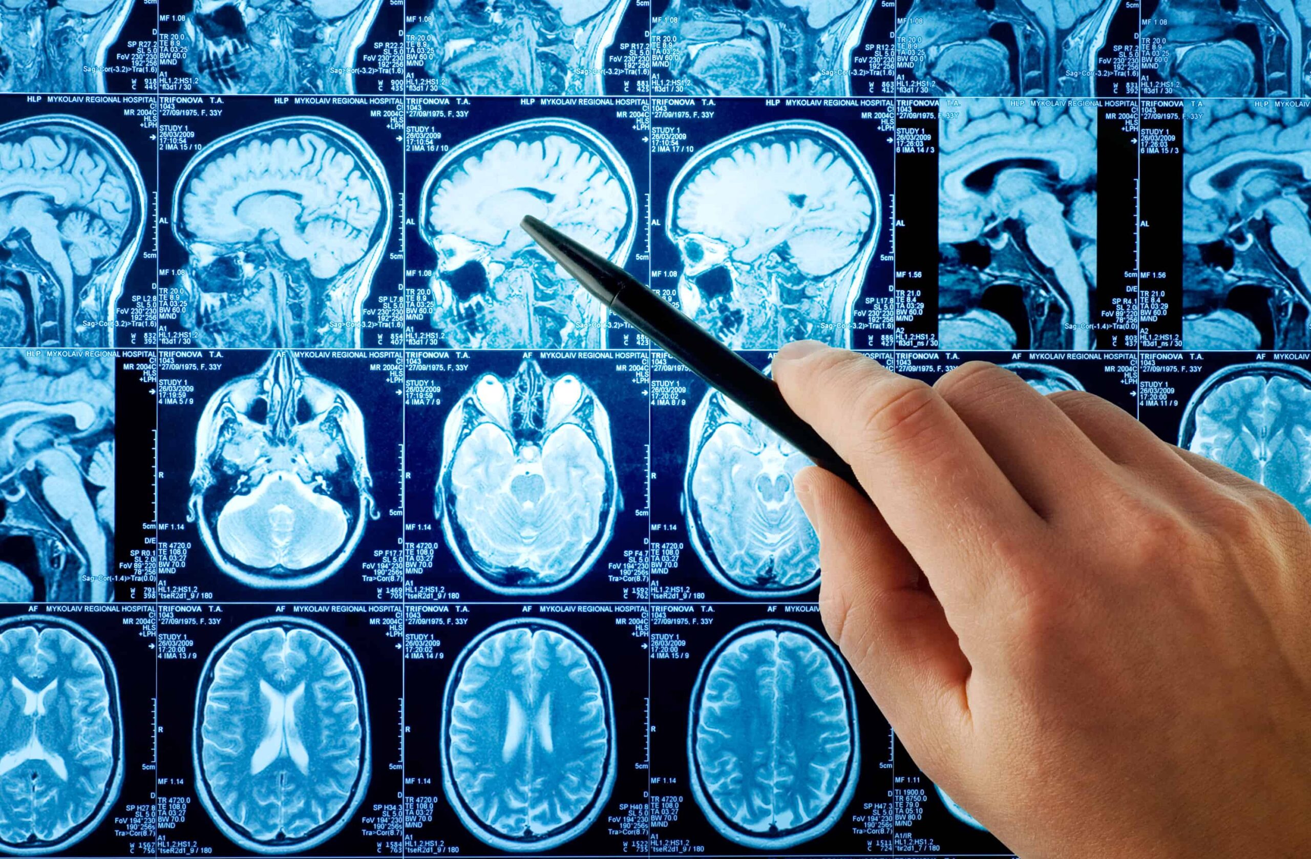
Q&A: Brain Imaging Research in Autism
Marvin Natowicz, MD, PhD, Rajesh Kana, PhD
The accredited educational series jointly provided by Autism Research Institute and Cleveland Clinic Center for Continuing Education reviews the co-occurrence…
Brain Imaging Research in Autism: Promises and Pitfalls
Marvin Natowicz, MD, PhD, Rajesh Kana, PhD
The accredited educational series jointly provided by Autism Research Institute and Cleveland Clinic Center for Continuing Education reviews the co-occurrence…

Helping Children with Autism Tune Into Voices: What Brain Science Reveals About Communication
Daniel Abrams, PhD
This talk explores how the brain processes speech and voices in autism, and how these differences relate to real-world communication.…